Labeled Picture Of Nasal Cavity : Human Nose Wikipedia : The nasal cavity is divided into equal halves by the nasal septum and the vomer bone.
on
Get link
Facebook
X
Pinterest
Email
Other Apps
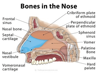
Labeled Picture Of Nasal Cavity : Human Nose Wikipedia : The nasal cavity is divided into equal halves by the nasal septum and the vomer bone.. Nasal cavity facts, function, parts and diseases, a comprehensive study. Shows the frontal, sphenoid and maxillary sinuses. The ridges on the nasal septum are enlarged, which can block airflow. The nasal cavity forms part of the aerodigestive tract. A pair of nasal bones support the bridge of the nose and the medial plates and frontal processes of the maxilla each image highlights and labels the sinuses in coronal and sagittal view.
The nasal septum divides the cavity into two cavities, also known as fossae. Each cavity is the continuation of one of the two nostrils. Polish your personal project or design with these nasal cavity transparent png images, make it even more personalized and more attractive. Nasal cavity model only, other accessories demo in the picture is not included. Gross anatomy the nasal cavity is formed by 1:
16 Nose Facts For Kids Students And Teachers from www.factsjustforkids.com Learn vocabulary, terms and more with flashcards, games and other study tools. The normal position of the nasal septum creates two roughly symmetrical nasal cavities. 34.1 skeleton of the nose the skeleton of the nose is composed of an upper bony portion and a lower cartilaginous portion. Check out our nasal cavity print selection for the very best in unique or custom, handmade pieces from our shops. Polish your personal project or design with these nasal cavity transparent png images, make it even more personalized and more attractive. The nasal cavity forms part of the aerodigestive tract. On the lateral nasal wall show the superior, middle and inferior nasal chonchae project medially into the nasal cavity forming the superior paranasal sinuses: Gross anatomy the nasal cavity is formed by 1:
Floor of the nasal cavity.
The nasal cavity is posterior to the nose and is framed and supported by several bones and cartilages. The horizontal plate of the palatine bone posteriorly and the palatine process of the maxilla anteriorly. To download a bluelink image, click on the photo and select the download option on the following page. In this article, we shall look at the applied anatomy of the nasal cavity, and some of the relevant clinical syndromes. Maxillary sinuses are in the cheek area, below the eyes on either side of the nose. From wikimedia commons, the free media repository. Bones of the nasal cavity. The nasal cavity is the most superior part of the respiratory tract. It consists of nasal skeleton, which houses the nasal cavity. Because most nasal cavity imaging for chronic sinusitis is currently performed with computed tomography, this article concentrates on ct understanding the anatomy of the nasal cavity and its anomalies is important because it leads to an understanding of imaging anatomy, which is needed to. Cribriform plate of the ethmoid. Along with the nose, paranasal sinuses and nasolacrimal duct, the nasal cavity forms the nasal. The photograph may be purchased as wall art, home decor illustration of the upper respiratory tract, nasal cavity.
Cribriform plate of the ethmoid. Bones of the nasal cavity. Atlas » cytology » nasal cavity. The nasal cavity is the most superior part of the respiratory tract. In this article, we shall look at the applied anatomy of the nasal cavity, and some of the relevant clinical syndromes.
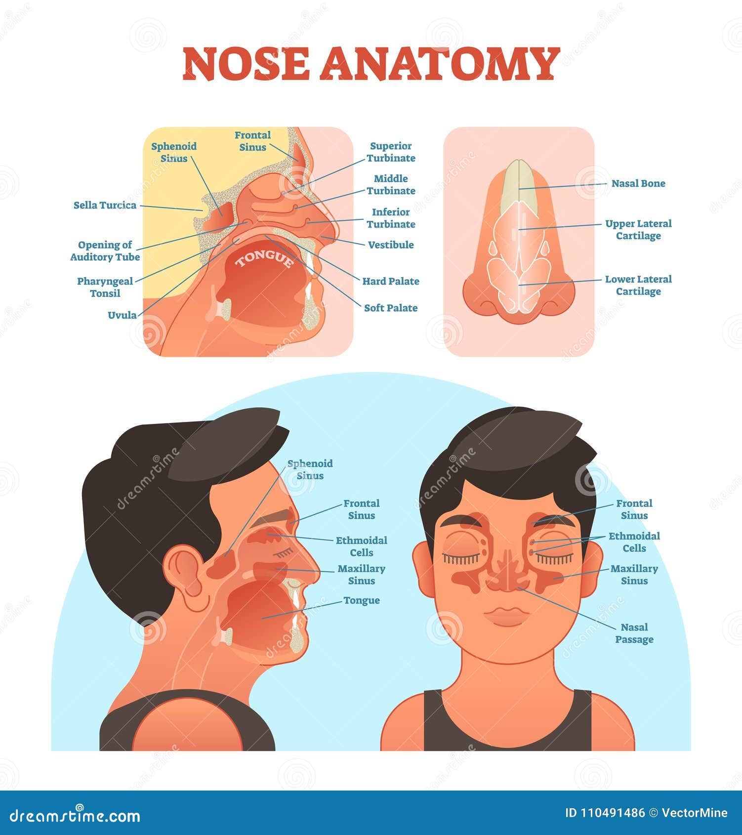
Nose Anatomy Medical Vector Illustration Diagram Stock Vector Illustration Of Nostril Mouth 110491486 from thumbs.dreamstime.com The photograph may be purchased as wall art, home decor illustration of the upper respiratory tract, nasal cavity. From wikimedia commons, the free media repository. Bones of the nasal cavity. The nasal cavity forms part of the aerodigestive tract. It consists of nasal skeleton, which houses the nasal cavity. To download a bluelink image, click on the photo and select the download option on the following page. Atlas » cytology » nasal cavity. Nasal cavity model only, other accessories demo in the picture is not included.
800 x 597 jpeg 68 кб.
800 x 597 jpeg 68 кб. Extreme lateral deviation of the septum may result in. Check out our nasal cavity print selection for the very best in unique or custom, handmade pieces from our shops. Nasal cavity model only, other accessories demo in the picture is not included. The nasal cavity is posterior to the nose and is framed and supported by several bones and cartilages. Clinical anatomy of the nose, nasal cavity and paranasal sinuses, thieme, chapter 2 nasal anatomy learning strategies. Each cavity is the continuation of one of the two nostrils. They can happen due to inflammation from asthma, chronic sinus infections, and nasal allergies (such as hay fever). Maxillary sinuses are in the cheek area, below the eyes on either side of the nose. Floor of the nasal cavity. Medial wall of nasal cavi… The nasal cavity conditions the air to be received by the other areas of the respiratory tract. The movement of air through.
The nasal cavity forms part of the aerodigestive tract. Nasal cavity definition, anatomy, functions, diagrams. The nasal cavity opens into a network of sinuses: Because most nasal cavity imaging for chronic sinusitis is currently performed with computed tomography, this article concentrates on ct understanding the anatomy of the nasal cavity and its anomalies is important because it leads to an understanding of imaging anatomy, which is needed to. The reserve crowns of the upper cheek teeth and a portion of the the epithelium of the nasal cavity changes from stratified squamous epithelium to pseudostratified columnar ciliated epithelium, composed of.
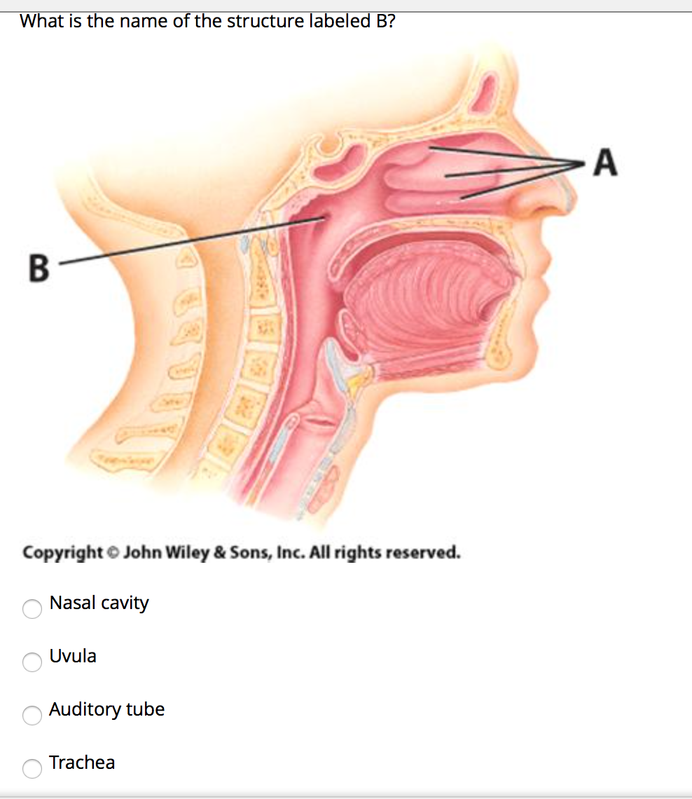
Solved What Is The Name Of The Structure Labeled B A B C Chegg Com from media.cheggcdn.com The photograph may be purchased as wall art, home decor illustration of the upper respiratory tract, nasal cavity. Jump to navigation jump to search. Polish your personal project or design with these nasal cavity transparent png images, make it even more personalized and more attractive. Atlas » cytology » nasal cavity. The nasal cavity conditions the air to be received by the other areas of the respiratory tract. Extreme lateral deviation of the septum may result in. The area just inside the nostrils together the paranasal sinuses and the nasal cavity filter and warm the air, and make it moist before it goes into the lungs. Along with the nose, paranasal sinuses and nasolacrimal duct, the nasal cavity forms the nasal.
The goal of the surgery is to remove the whole tumor and a small amount of normal.
Here you can explore hq nasal cavity transparent illustrations, icons and clipart with filter setting like size, type, color etc. The nasal cavity conditions the air to be received by the other areas of the respiratory tract. Labeled nasal cavity, sinuses and ear images. The nasal cavity forms part of the aerodigestive tract. Colour illustration of the anatomy of the human nasal, oral and laryngeal cavitites and related structures (sagittal section). The ridges on the nasal septum are enlarged, which can block airflow. Nasal cavity model only, other accessories demo in the picture is not included. Polish your personal project or design with these nasal cavity transparent png images, make it even more personalized and more attractive. Medial wall of nasal cavi… 34.1 skeleton of the nose the skeleton of the nose is composed of an upper bony portion and a lower cartilaginous portion. The nasal septum divides the cavity into two cavities, also known as fossae. Medial wall of nasal cavity (nasal septum). Maxillary sinuses are in the cheek area, below the eyes on either side of the nose.
Each cavity is the continuation of one of the two nostrils nasal cavity labeled. Cribriform plate of the ethmoid.
Comments
Post a Comment